Importantly, 27 of 30 (90%) cancers reported by Berg et al ( 5) were ductal carcinoma in situ (DCIS) of the 25 cancers with grading, 15 (60%) were low-nuclear-grade DCIS, and all three invasive malignancies were low grade. Subsequently, Burnside et al ( 6) reported a malignancy rate of four of 30 (13%) for biopsied amorphous calcifications, and Bent et al ( 7) showed 10 of 51 (20%) lesions were malignant. Since that time, such calcifications have generally been assessed as suspicious (Breast Imaging Reporting and Data System category 4b), and recommended for biopsy ( 2, 4, 5).
Breast calcification clusters series#
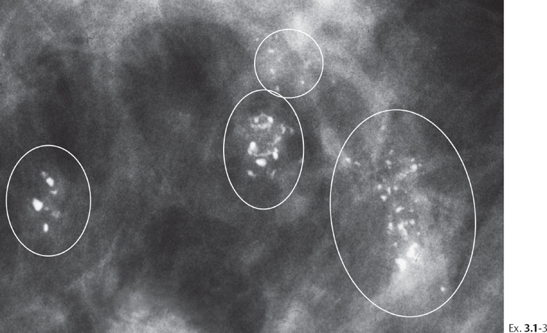
In 2001, in a prospective series of 150 patients in which all amorphous calcifications (other than those scattered in both breasts) were recommended for biopsy, 30 of 150 (20%) patients had malignant lesions ( 5). In 1998, Liberman et al ( 4) reported that nine of 35 (26%) biopsied amorphous calcifications were malignant. Historically, management of amorphous calcifications varied from surveillance to biopsy ( 3). Amorphous calcifications are indistinct, appearing so small or hazy even on spot magnification mammograms that more specific morphologic classification cannot be assigned ( 2). While the majority of calcifications seen at screening mammography can be dismissed as typically benign, calcifications can be the earliest detectable evidence of breast cancer 55% of nonpalpable breast cancers have associated calcifications at mammography ( 1). Mammographically detected breast calcifications are analyzed based on morphology and distribution. In women younger than 50 years without a personal history of cancer, grouped amorphous calcifications showed four of 127 (3.1%) (95% CI: 0.9, 7.9) were malignant and 39 of 127 (30.7%) were atypical at final histopathology. Of 356 grouped amorphous calcifications, 102 (28.7%) yielded atypical results prompting excision, with three of 102 (2.9%) upgraded to ductal carcinoma in situ at excision. Malignancy rates in a segmental (six of 21 ), linear (eight of 32 ), or multiple group same quadrant (nine of 36 ) distribution were significantly higher than malignancy rate in a solitary group of amorphous calcifications (25 of 356 ) ( P =. Fifty-two (10.5% 95% confidence interval : 7.9, 13.5) lesions proved malignant, with 17 of 52 (42.7%) being invasive cancers (median, 0.3 cm range, 0.1–1.3 cm) and all 17 of them being estrogen and progesterone receptor positive and node negative.

After excluding atypical lesions not excised and patients with more than one biopsy in the same year, 497 lesions from 494 women (median age, 52 years age range, 30–81 years) remained. Of 1903 sequential biopsies, 546 (28.7%) were for amorphous calcifications.


 0 kommentar(er)
